
Presentation
Dyspnea
Patient Data


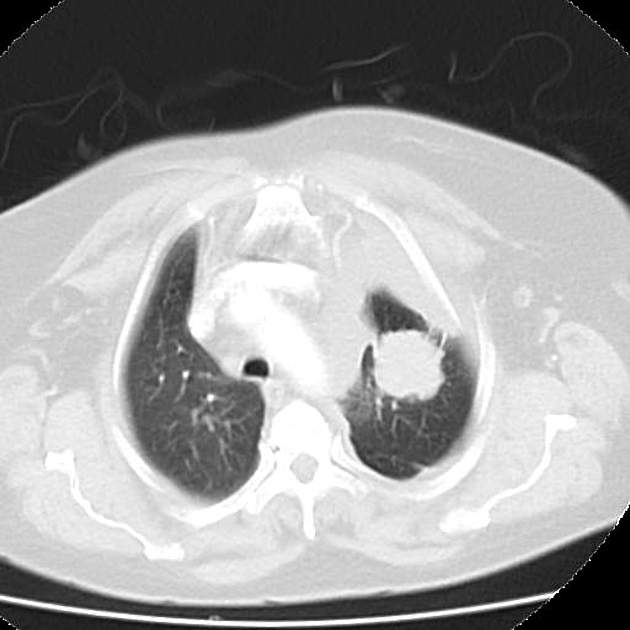
A fairly well-defined, roughly spherical pulmonary mass lesion is seen in the left upper lobe. It measures about 3.7 x 3.5 cm along its maximum axial diameters. It shows a spiculated margin with no evidence of matrix calcification or internal breakdown. The lung parenchyma around the lesion showed fine reticulations with a pleural tail connecting the mass to the lateral chest wall.
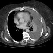
Marked left sided pleural effusion with subsequent subtotal compression collapse of the left lung and contralateral shifting of the trachea and mediastinum.
Subsegmental atlectatic bands are seen at the middle lobe, otherwise clear right lung field.
Mildly enlarged (about 1.4 cm) prevasuclar and retrocaval lymph nodes are noted.
Multiple rounded small osteolytic bone lesions are seen involving the dorsal vertebra.
Case Discussion
Left upper lobar pulmonary mass with spiculated margin: bronchogenic carcinoma with mild regional mediastinal lymphadenopathy and left sided marked pleural effusion.


 Unable to process the form. Check for errors and try again.