
Presentation
Left maxillary swelling.
Patient Data
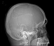

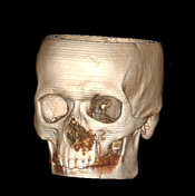

diffuse bone sclerosis involving the skull bones, mandible and scanned cervical vertebrae with widespread minute lytic lesions seen scattered within giving the skull the characteristic salt and pepper appearance
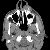
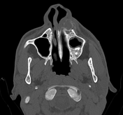
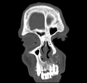
an expansile multilocular osteolytic lesion is seen at the left side of the superior alveolar margin. No associated cortical destruction, periosteal reaction or extra-osseous soft tissue component
mild mucosal thickening of the left maxillary sinus obliterating the left osteomeatal unit
mild mucosal thickening of the frontal sinus as well as the anterior ethmoid air cells
bilateral middle concha bullosa
mild rightward deviation of the nasal septum
Case Discussion
The patient had a history of chronic renal failure and on dialysis. He presented with left maxillary swelling.
CT findings are in keeping with renal osteodystrophy with left maxillary brown tumor.


 Unable to process the form. Check for errors and try again.