
Presentation
Acute abdominal pain.
Patient Data


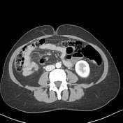

There is an abnormal position of the cecum which passes through the foramen of Winslow to lie within the lesser sac. The foramen of Winslow is widened with the main portal vein partially effaced by the adjacent large bowel. Both the terminal ileum and appendix have a normal appearance with the appendix lying outside of the lesser sac. The cecum measures up to 4.3 cm. No small bowel obstruction.
Ill-defined hypodensity adjacent to the falciform ligament likely represents a region of focal fatty infiltration. No other focal liver lesion identified. Nonspecific mild periportal edema. No dilatation of the common bile duct. The pancreas, spleen, right kidney and adrenals are unremarkable. Calcified density within/adjacent to the left adrenal gland noted. Peripheral hypodensities within the mid and lower pole left kidney are too small to accurately characterize on CT but likely represents small cysts.
There is a small volume of free intra-vesical gas, with no IDC and preservation of the fat plane around the bladder. 3.4 cm ovoid hypodense right adnexal lesion.
Small partly imaged peri-fissural nodule associated with the anterior right oblique fissure.
Conclusion:
Internal herniation of the cecum into the lesser sac, with the cecum measuring up to 4.3 cm. Normal appearance of the appendix which lies predominantly outside the lesser sac. No bowel dilatation identified at the time of study.
Intravesical gas, due to recent catheterization.
3.4 cm hypodense right adnexal lesion likely represents a cyst, for ultrasound correlation if clinically warranted
Case Discussion
There is an internal hernia, the cecum lies in the lesser sac having herniated through the foramen of Winslow.


 Unable to process the form. Check for errors and try again.