
Presentation
Acute chest pain and dyspnea.
Patient Data

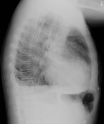
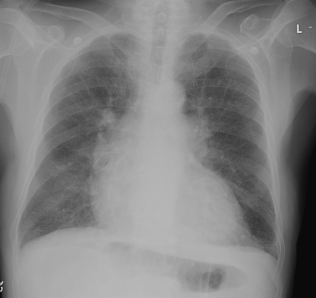
Bilateral perihilar opacification, upper lobe vascular redistribution and increased interstitial markings in keeping with acute pulmonary edema. Bilateral posterior pleural effusions on the lateral projection.
Cardiomegaly. A curvilinear calcific density is projected over the left ventricle outlining the cardiac chamber.
Case Discussion
Mural thrombus in the left ventricle can form following myocardial infarction where there is a loss of normal myocardial contractility, causing to stasis and thrombosis. Chronic mural thrombus can subsequently calcify as is the case in this man. It is also associated with ventricular aneurysm which was not identified on echocardiography in this patient.
This patient had a history of multiple prior acute myocardial infarctions. He was treated for pulmonary edema due to worsening cardiac function.


 Unable to process the form. Check for errors and try again.