
Presentation
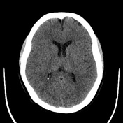
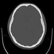
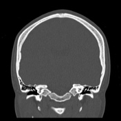
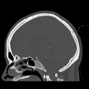
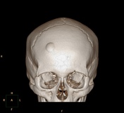
Longstanding lump in right frontal region.
Patient Data
Age: 20 years
Gender: Female

Download
Info

Simple exostosis of 5.8 x 23.3 mm in the right frontal region. Unenhanced CT of the brain with no other relevant pathological findings.
Case Discussion
This case demonstrates a simple frontal osteoma.


 Unable to process the form. Check for errors and try again.