Presentation
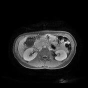
Known case of cervix carcinoma. Follow-up imaging after 1 year.
Patient Data
Age: 55 years
Gender: Female
From the case:
Carcinoma of the cervix - recurrence















Download
Info
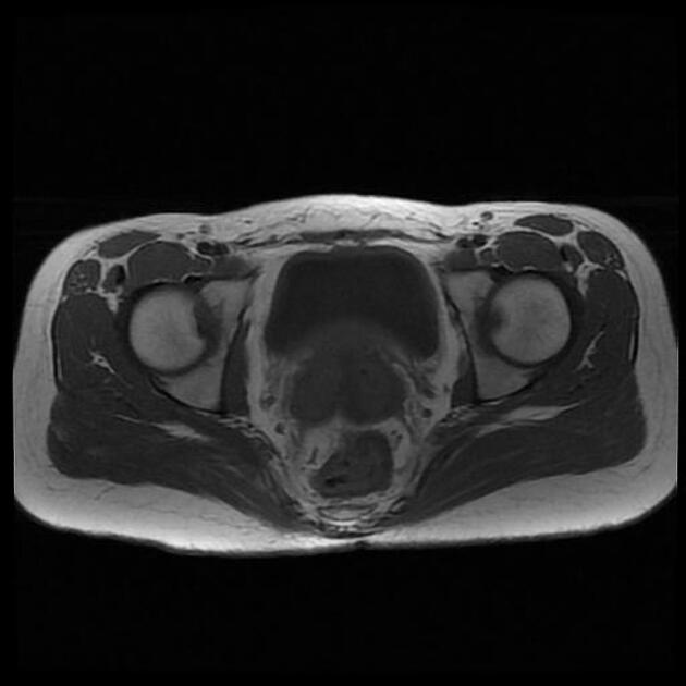
TAH+BSO.
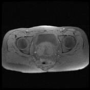
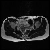
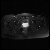
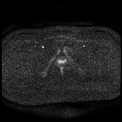
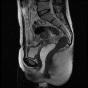
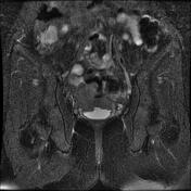
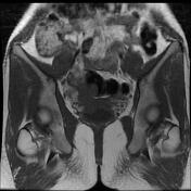
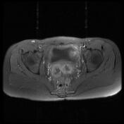
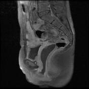
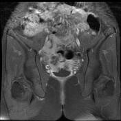
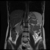
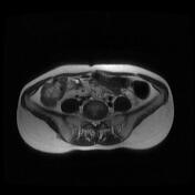
Two abnormal signal solid necrotic enhancing infiltrative mass lesions measuring about 48 x 50 mm and 20 x 28 mm with marked water restriction on DWI images at the site of surgical resection just above the vaginal cuff that shows invasion to :
- vaginal cuff
- serosal surface of urinary bladder posterior wall
- mesorectal fascia and the serosal surface of the sigmoid and ileal loops
Case Discussion
Recurrent carcinoma of the cervix.


 Unable to process the form. Check for errors and try again.