
Presentation
Slow atrial fibrillation. Cardiomyopathy on echocardiogram.
Patient Data
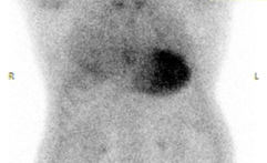
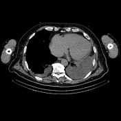


There is abnormal increased uptake of tracer in the myocardium (most avid in the left ventricle) at one hour. Anatomical localization confirmed on the fused SPECT-CT.
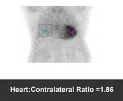
Cardiac uptake is much greater than rib uptake, with relative suppression of tracer accumulation in the ribs (Grade 3). Heart to contralateral thorax ratio is elevated (1.86, normal less than 1.5). The scan is considered positive for ATTR amyloidosis given the clinical history.
Cardiomegaly. Small pericardial effusion. Bilateral pleural effusions (left greater than right) with adjacent basal collapse.
Case Discussion
This case demonstrates the typical imaging appearances of ATTR cardiac amyloidosis on nuclear medicine amyloidosis study. The combination of planar imaging and SPECT-CT improves the anatomical localization of the abnormal tracer uptake in these studies.
Fused SPECT-CT images are particularly useful in cases where there is less intense accumulation of tracer in the heart. In these cases, SPECT-CT allows distinction of physiological blood pool avidity and abnormal myocardial uptake.


 Unable to process the form. Check for errors and try again.