
Presentation
Left proptosis for 2 weeks.
Patient Data
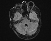

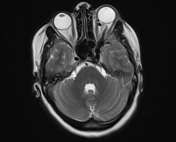
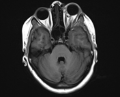
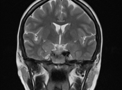
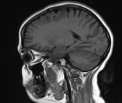
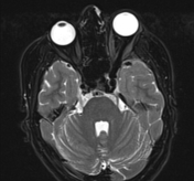
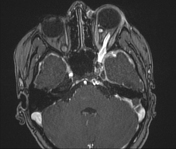
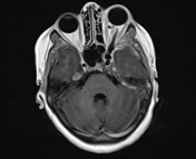
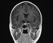
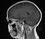

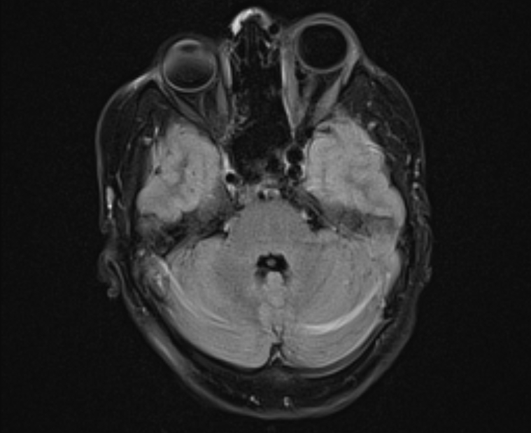
Mild proptosis of the left eye globe associated with hugely dilated superior ophthalmic vein anatomizing with dilated preseptal and superficial veins of the face.
Engorged left cavernous sinus vessels with suspected connection between the cavernous portion of the left internal carotid artery and the cavernous sinus. Enlarged inferior petrosal sinus and thickened left temporal dural reflection.
Case Discussion
Features are suggestive of left direct caroticocavernous fistula. Presentation is usually with pulsatile exophthalmos. Dilatation of superior ophthalmic vein and engorgement of the cavernous sinus are key imaging features of caroticocavernous fistula in cross-sectional imaging. Angiography is the gold standard for the diagnosis, type classification and planning for embolization.


 Unable to process the form. Check for errors and try again.