
Presentation
Nontender swelling in the right level 2 cervical region for 3 years.
Patient Data
Age: 25 years
Gender: Female
From the case:
Carotid body tumor

Download
Info

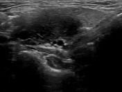
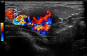
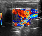
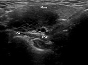
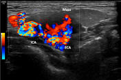
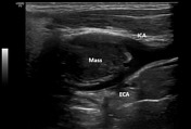
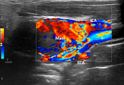
A (38 x 24 x 11 mm) well-defined heterogeneously hypoechoic mass is seen in right side level 2 cervical regions at the carotid bifurcation which is splaying the Internal and external carotid artery. On color Doppler study the mass is taking intense vascularity.
Case Discussion
Final Diagnosis: Carotid body tumor
Ultrasonography: Round to oval well define heterogeneously hypoechoic mass in the lateral neck with splaying of the common carotid artery. Small vessels can be demonstrated within the mass
Differential diagnosis: Nerve sheath tumors, nodal metastasis, abscess, vascular thrombosis.
Rarely lipoma, liposarcoma, hibernoma.
Treatment: Surgical excision.


 Unable to process the form. Check for errors and try again.