
Presentation
Thumb, index and middle finger parasthesia.
Patient Data
Age: 60 years
Gender: Male
From the case:
Carpal tunnel syndrome

Download
Info

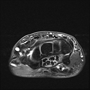
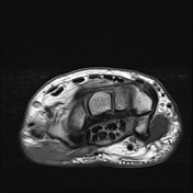
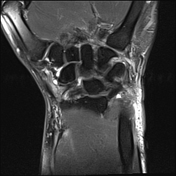
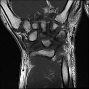
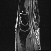
Edema within the carpal tunnel. The median nerve is flattened within the carpal tunnel at the level of the hook of hamate with thickening and palmar bowing of the flexor retinaculum. Proximally, the median nerve is high signal and appears swollen with a cross-sectional diameter of 30 mm2 at the level of the pisiform. Overlying subcutaneous edema.
Case Discussion
Typical MRI features of carpal tunnel syndrome:
- palmar bowing of the flexor retinaculum
- nerve thickening and edema at the carpal tunnel inlet (level of the pisiform) with increased cross-sectional area
- nerve flattening at the carpal tunnel outlet (level of the hook of hamate)
- edema within the carpal tunnel


 Unable to process the form. Check for errors and try again.