
Presentation
Treated for ovarian cancer on follow-up since 4 years. Incidental finding on chest CT.
Patient Data
Age: 60 years
Gender: Female
From the case:
Catheter pulmonary embolism

Download
Info
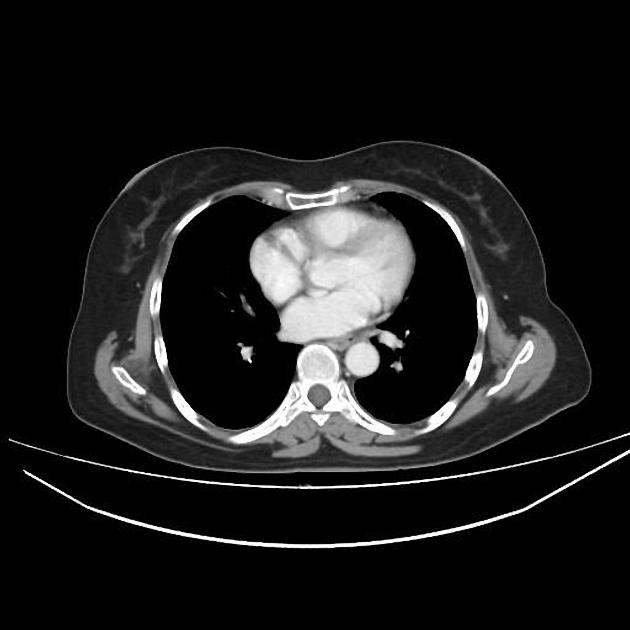
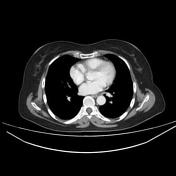
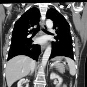
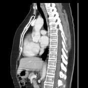
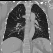
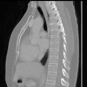
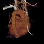
A fractured catheter fragment is seen in the segmental and subsegmental posterior basal branches of the right lower pulmonary artery.
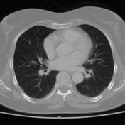
Both lungs are clear. No pleural effusion is seen.
No mediastinal or hilar lymphadenopathy
Case Discussion
CT features of a catheter pulmonary embolism seen as fractured catheter fragment in the segmental and subsegmental posterior basal branches of the right lower pulmonary artery.
This patient had a central venous catheter in the subclavian vein during her treatment with chemotherapy.
For the main complications of the central venous catheter (CVC), please refer to the article: central venous catheter.


 Unable to process the form. Check for errors and try again.