
Presentation
Right lower quadrant pain.
Patient Data
Age: 35 years
Gender: Female
From the case:
Cecal diverticulitis

Show annotations
Download
Info

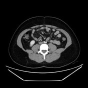
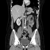
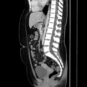
Single inflamed diverticulum projecting from the medial cecum with surrounding fat stranding, reactive submucosal edema of the cecum and minor inflammation of the terminal ileum. Few smaller noninflamed diverticula. Trace ascites. Normal appendix.
Case Discussion
Very clear imaging presentation of cecal diverticulitis which serves as a good reminder that diverticulitis can occur throughout the GI tract from the duodenum through the sigmoid colon. In this case, cecal diverticulitis is a clinical mimic for appendicitis.


 Unable to process the form. Check for errors and try again.