
Presentation
Abdominal cramps and vomiting for a few hours.
Patient Data
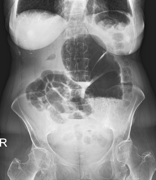
A gross distended large bowel loop (note haustrations) is seen slightly left of the midline, its "hilum" is oriented towards the right.
Dilated small bowel loops in the right lower quadrant.
The descending colon and splenic flexure is to the left of this loop and has normal appearance.
Altogether, findings are highly suspicious for cecal volvulus.
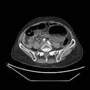


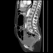
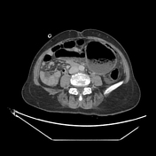

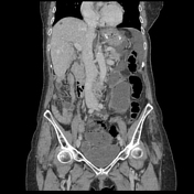

CT confirms the cecal volvulus, note the well depicted whirlpool sign in the sagittal plane at the level of the aortic bifurcation.
Other: moderate free fluid, small calcific pelvic lesion on the left (unchanged compared to old prior exam), hepatic cysts, cysts and kidney stones, mild common bile duct dilation, small umbilical hernia, nasogastric tube in situ, degenerative skeletal changes.
Case Discussion
During surgery the cecal volvulus was confirmed, manual detorquation was first performed, which however did not relieve the small bowel ileus, thus a right hemicolectomy was performed.
Histopathological assessment of the resected specimen showed ischemic changes of the colonic wall, but no sign of malignancy.


 Unable to process the form. Check for errors and try again.