
Presentation
Characterization of a palpable lump in her palate, on the right side.
Patient Data
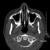

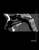

There is a solitary 2.1 x 1.8 cm expansile lucent lesion in the alveolar process of the right maxilla. There is no associated impacted tooth although it appears to abut the apical regions of the ipsilateral premolar and molar teeth. There is an apparent breach of the cortex into the nasal cavity, best appreciated on the sagittal reformatting.
Differential diagnosis include, central giant cell granuloma, odontogenic keratocyst and less likely ameloblastoma.
Case Discussion
Histology:
Fragments consist of cellular proliferation with a conspicuous population of multincelate giant cells. Between the giant cells are monocluate variants with similar nuclear profiles. There is a further population of bland, spindled, fibroblasts-like cells with associated hemosiderin and scattered inflammation. Rare fragments of reactive bone are also seen. No atypical mitoses are appreciated.
The findings are consistent with central giant cell granuloma.


 Unable to process the form. Check for errors and try again.