
Note: This case has been tagged as "legacy" as it no longer meets image preparation and/or other case publication guidelines.
From the case:
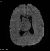
Central neurocytoma


Download
Info
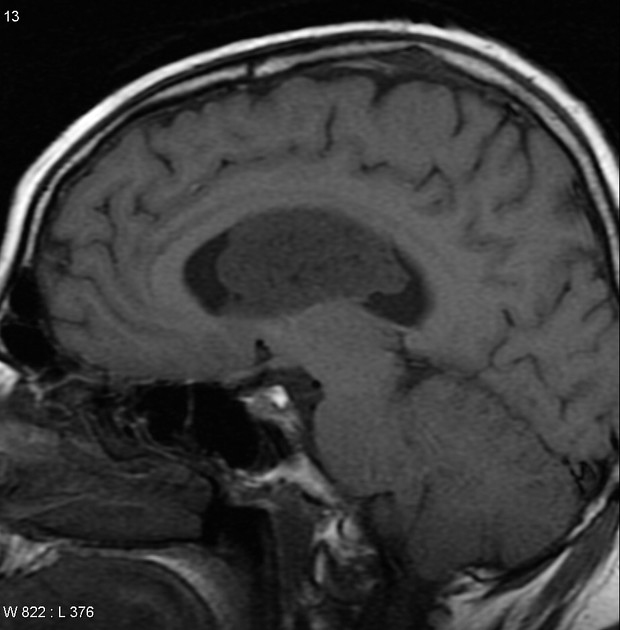
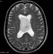
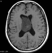
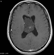
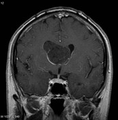
MRI of the brain including post contrast sequences demonstrates a large intraventricular mass filling and expanding the body of the right lateral ventricle. It has high T2 signal, low T1 signal and has multiple small cystic regions within it (bubble appearance). Following contrast administration only minimal heterogeneous enhancement is present.
Case Discussion
This case illustrates typical appearances of a central neurocytoma, confirmed histologically.


 Unable to process the form. Check for errors and try again.