
Presentation
Worsening headaches.
Patient Data
Age: 25 years
Gender: Male
From the case:
Central neurocytoma


Download
Info
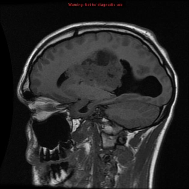
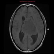
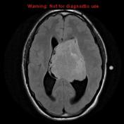
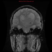
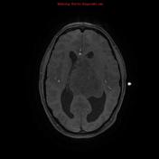
There is a heterogeneously enhancing intraventricular mass with focal intralesional low T1/high T2 signal, which could represent either cystic spaces or necrosis.
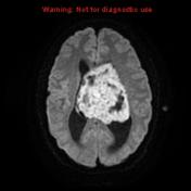
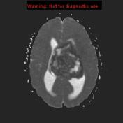
There is evidence of restricted diffusion. Patchy areas of susceptibility may be due to either calcification or blood products.
The lateral ventricles are enlarged and there is distortion and displacement of the septum.
The patient went on to have surgery which confirmed a central neurocytoma.
Case Discussion
This case demonstrates typical appearances of a large central neurocytoma.


 Unable to process the form. Check for errors and try again.