
Presentation
Fever, headache, and vomiting for the past week. History of recurrent otitis media. CT performed to assess for complications of otitis media.
Patient Data
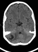


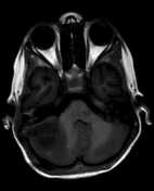
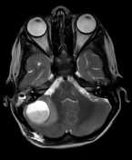
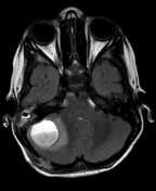
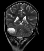
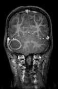
Round intra-axial lesion with a diameter of 2.4 cm in the right cerebellar hemisphere, containing mixed contents and displaying ring enhancement after contrast injection. Perilesional edema with mass effect manifesting as narrowing of the fourth ventricle on the right. The midline is preserved.
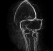
No evidence of venous sinus thrombosis.
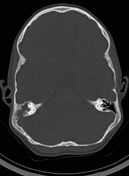
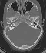
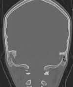
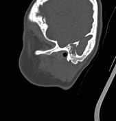
Total opacification of the right middle ear and mastoid air cells, with erosion of the posterior mastoid wall.
In summary: Right otomastoiditis with posterior mastoid wall erosion, resulting in abscess formation in the right cerebellar hemisphere.

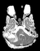

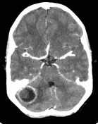
Status post right occipital craniotomy and mastoidectomy. Signs of hemorrhage in the surgical tracks.
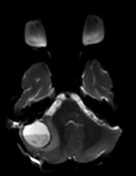
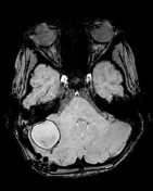
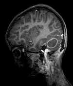
Right cerebellar abscess measuring 3.0 x 2.6 x 2.5 cm, with a thick, enhancing wall, containing pus with a level and a small amount of blood. The right and superior walls of the abscess abut the tentorium cerebelli. The tentorium is mildly thickened. Perilesional edema.
The right cerebellar tonsil is prolapsed 4 mm below the foramen magnum. The fourth ventricle is compressed on the right.
In the surgical space in the right ear there is an opacity with signs of blood but no collection of pus.
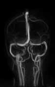
No thrombus identified on MRV in the main dural venous sinuses.
In summary: Right cerebellar abscess, with no significant change.
Case Discussion
The cerebellar abscess shrank substantially in size on a subsequent MRI scan done a month later (not shown).


 Unable to process the form. Check for errors and try again.