
Presentation
Acute onset of aphasia and right paraplegia.
Patient Data
Age: 22 years
Gender: Female
From the case:
Cerebral sinus venous thrombosis with lobar hemorrhage
Download
Info
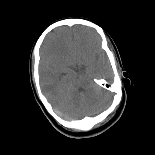
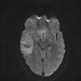
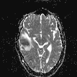
Acute lobar hematoma (2.9 x 2.1 cm) is seen in the right temporo-occipital region with surrounding edema.
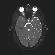
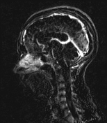
The hyperdense appearance of the superior sagittal sinus and right transverse and sigmoid sinuses is noted.
The rest of the brain parenchyma is normal.
From the case:
Cerebral sinus venous thrombosis with lobar hemorrhage

Download
Info

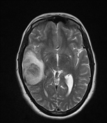
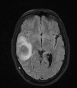
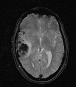
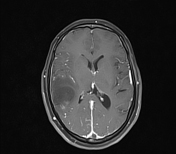
Acute lobar hematoma with surrounding edema in the right temporoparietal region.
Extensive filling defects involving the cerebral superficial cortical veins, superior sagittal sinus extending to the torcular herophili, right transverse sinus, and right sigmoid sinus.
Case Discussion
Cerebral venous sinus thrombosis is a well-known cause of lobar hemorrhage in young patients.


 Unable to process the form. Check for errors and try again.