
Presentation
Postural neck pain and sensations of intracranial pressure, especially when standing up from a lying position, since several months. No recent trauma. Relapsing-remitting multiple sclerosis since 17 years, treated with intrathecal triamcinolone in past episodes. No recent lumbar puncture.
Patient Data


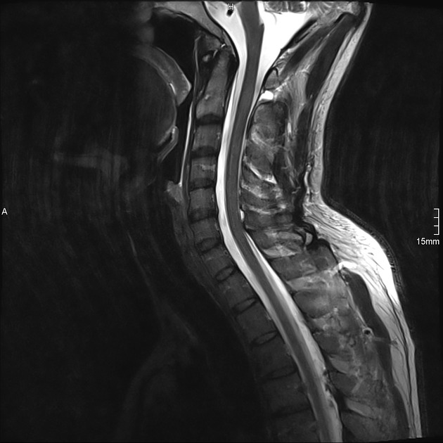
There is an extensive extradural fluid collection separating the dura from the bony spinal canal (curtain sign).
The extradural fluid collection shows a T2 hyperintense signal compared to CSF (which shows a slightly lower signal due to flow/pulsation artifacts). The fluid collection shows a T1 hyperintense signal to CSF, probably due to proteinaceous content. The differential diagnosis of an epidural hemorrhage is excluded by a sagittal haemosensitive sequence which shows no blood products. We therefore suspect a spontaneous cerebrospinal fluid leak (CSF leak) with intracranial hypotension as a cause for the patients postural symptoms.
The cranial T1 C+ sequence demonstrates diffuse smooth enhancing pachymeningeal thickening as well as the venous distension sign (rounding of the dominant dural venous sinus in sagittal imaging) fortifying the diagnosis of intracranial hypotension.


CT post myelogram confirms an extradural collection of contrast enhanced cerebrospinal fluid and suggests that the location of the leak into the epidural space lies dorsally at the height of C1/2.
It is critical to recognize that contrast at this level is sometimes a false localizing sign as contrast ascends the in epidural space to the C1/2 level.
Nonetheless, given that the contrast is most concentrated at this level, and little contrast is seen elsewhere in the epidural space, the cervical dural leak is treated by a CT-guided blood patch as demonstrated above.
Comparison of normal Cx...


Comparison of normal Cx spine same patient 2 years ago vs CSF leak
Comparison of normal cervical spine (same patient, 2 years ago) vs. CSF leak spine (same patient upon presentation).
Case Discussion
A patient's MRI with postural symptoms demonstrated:
an extensive extradural fluid collection separating the dura from the bony spinal canal (curtain sign) in spinal MRI and
signs of intracranial hypotension in cranial MRI (smooth enhancing pachymeningeal thickening, venous distension sign)
A post-myelogram CT demonstrates a cervical dural leak emitting contrast into the epidural space dorsally at the height of C1/2.
Important: It should be noted that an extradural contrast collection at C1/2 is often the case in patients with extensive epidural leakage because the leaked fluid can migrate significant distances and pool depending on patient position and anatomy, and it does not, in such cases, mean that the leak is definitely located at C1/2. This is known as the C1-C2 false localizing sign. In this case, however, the paucity of contrast in the epidural space elsewhere is more suggestive that this is actually the site of the leak.
Therefore we concluded the diagnosis as cervical CSF leak leading to intracranial hypotension with postural symptoms.
The leak was later treated by a CT-guided blood patch at the C1/2 level. The patient did not show any postural symptoms from the day after treatment.


 Unable to process the form. Check for errors and try again.