
Presentation
Palpable mass on the anterior chest wall. History of mediastinal radiotherapy due to Hodgkin lymphoma.
Patient Data
Age: 80 years
Gender: Male
From the case:
Chest wall osteosarcoma


Download
Info
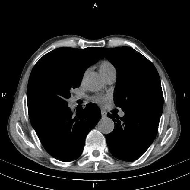
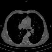
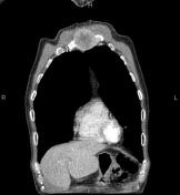
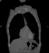
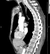
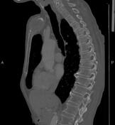
A 55×40 mm destructive mass is noted at the manubrium of the sternum that shows inhomogeneous enhancement on post contrast images. The mass infiltrates adjacent soft tissue structures and bulges into the superior mediastinum.
A 10mm hypodense lesion is noted in imaged portions of the upper abdomen at the liver.
Case Discussion
Pathology proved sternal osteosarcoma, a relatively rare entity, most often seen in elderly patients, mainly due to previous radiation therapy.


 Unable to process the form. Check for errors and try again.