
Presentation
Frequent headache, no neurologic deficit. No history of developmental delay.
Patient Data


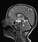
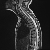
There is a caudal descent of the cerebellar peg-like tonsils about 15 mm through the foramen magnum (below McRae line) which appears crowded. The inferior tip of the herniated tonsil seen at the level of the lower endplate of the C2 vertebral body.
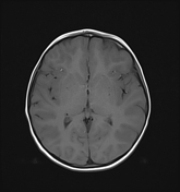
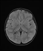
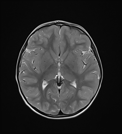
The CSF spaces are of normal size. No hydrocephalus. No shift of midline. No infarction or bleeding.
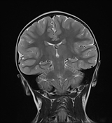
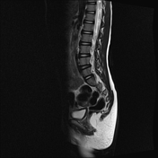
No intrinsic cord pathology. No syringohydromyelia. No tethered cord or spinal myelomeningocele. No vertebral body anomaly.
Case Discussion
Chiari I is the mildest and most common form of Chiari malformations recognized by the descent of cerebellar tonsil below the level of the foramen magnum. Most cases are asymptomatic, symptomatic patients complaining of headache, and neck pain. Up to 5 mm in children and 3 mm in adult tonsillar descent is considered normal.
A definitive diagnosis is an MRI study that shows tonsil descent measures on sagittal T1 or T2 weighted images. The presence of symptoms is directly related to the degree of cerebellar descent. Also, the presence of kink at the cervico-medullary junction is associated with a higher likelihood of developing symptoms.
The most common complication of Chiari I is syringomyelia seen in 25-50% of patients and most commonly seen in the cervical cord. Other less common complications include hydrocephalus.


 Unable to process the form. Check for errors and try again.