
Presentation
Painful left knee.
Patient Data
Age: 80 years
Gender: Male
From the case:
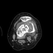
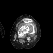
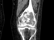
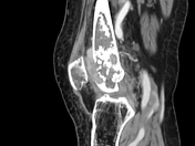
Chondrosarcoma - femur

Download
Info

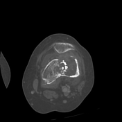
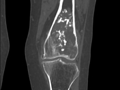
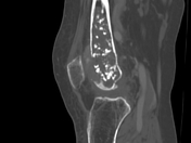
There is a large osteolytic lesion of the distal femur with enhanced tumoral matrix, containing rings and arcs calcification (cartilaginous matrix) with endosteal scalloping and cortical breach at several levels (mainly anterior). A soft tissue extension is noted mainly towards the lateral femoropatellar joint. A mild joint effusion is noted.
Case Discussion
CT features highly suggestive of a chondrosarcoma


 Unable to process the form. Check for errors and try again.