
Presentation
Headaches.
Patient Data
Age: 55 years
Gender: Male
From the case:
Chondrosarcoma - sphenoid wing

Download
Info
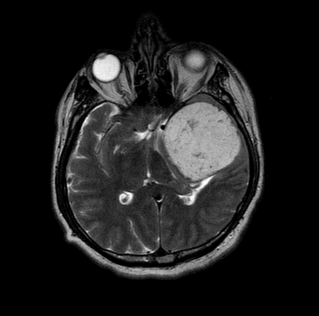
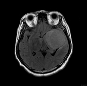
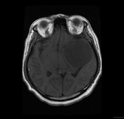
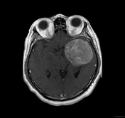
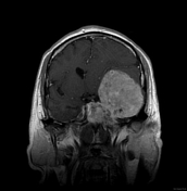
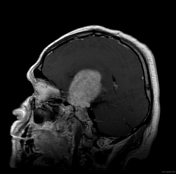
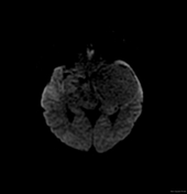
Selected MRI images demonstrate a large left sphenoid wing extra-axial mass that has very high T2 signal and vivid contrast enhancement. The adjacent brain is displaced with minimal edema.
The off-midline location and very high T2 signal suggest the diagnosis of chondrosarcoma.
Case Discussion
The patient went on to have a resection. Histology confirmed a chondrosarcoma.


 Unable to process the form. Check for errors and try again.