
Patient Data

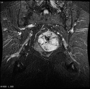
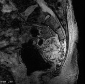

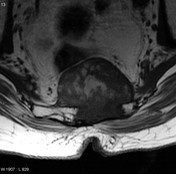
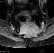
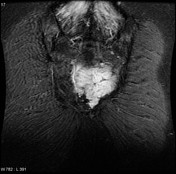

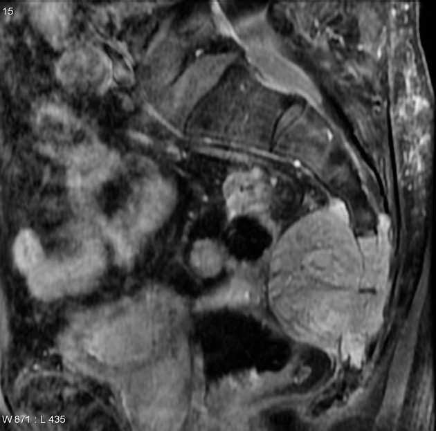
Heterogenous mass arising from the sacral vertebra, seen as predominantly hypointense on T1 and hyperintense on T2 with vivid contrast enhancement. Multiple non-enhancing hypointense areas represent matrix calcifications.
Case Discussion
The patient went on to have a biopsy.
Histology
The sections show strands and clusters of atypical epithelioid cells within a myxoid matrix. The tumor cells have oval to round, occasionally spindled, hyperchromatic nuclei with occasional larger nuclei with irregular contours. The cells have a moderate amount of vacuolated eosinophilic cytoplasm. Well developed physaliferous cells are not found. An occasional mitotic figure is found. There is no necrosis. Overt cartilaginous or osteoid tissue is not found.
By immunohistochemistry, the tumor cells are strongly positive for keratin. They are positive for S100 and EMA. They are negative for PSA.
FINAL DIAGNOSIS: chordoma


 Unable to process the form. Check for errors and try again.