
Presentation
Work up for prolonged intermittent abdominal pain.
Patient Data
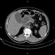


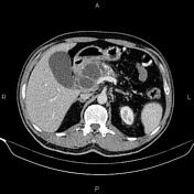
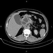

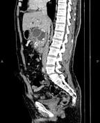

The pancreas is enlarged and contains several adjacent non-enhanced cystic lesions with a maximum diameter of 38 mm and a total diameter of 80 mm, particularly in the proximal portion. The pancreatic duct is dilated, and multiple coarse calcified foci are evident in the remnant parenchyma of the pancreas.
Intra and extra hepatic bile ducts are dilated, and CBD is measured 13 mm in diameter in the proximal portion.
The prostate gland is enlarged.
Case Discussion
The patient went on to have endoscopic ultrasonography and FNA.
Histology
Received 29 unstained slides prepared and stained with the Pap method.
Smears are cellularly composed of clusters of acinic cells and some ductal clusters mixed by a few islet cells in the degenerative background. No mucin was seen. Few pieces of fibrosis were noted.
Final diagnosis
Chronic pancreatitis
Negative for malignancy. No mucin.
No plasma cell representative of autoimmune pancreatitis was seen.
CT features of chronic pancreatitis include dilatation of the main pancreatic duct, pancreatic calcification, changes in pancreatic size, shape, and contour and pancreatic pseudocysts.


 Unable to process the form. Check for errors and try again.