
Presentation
Impaired renal function. Right hydronephrosis on ultrasound scan.
Patient Data
Age: 55 years
Gender: Female
From the case:
Chronic pelviureteric junction obstruction

Download
Info

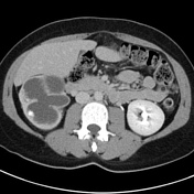
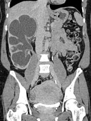
Severe right-sided hydronephrosis to the level of the pelviureteric junction. Ureter normal in caliber.
Advanced cortical thinning of the right kidney to 3 mm.
No meaningful excretion in the right collection system.
Normal left kidney.
Case Discussion
A late presentation of a pelviureteric junction obstruction. Abrupt caliber change at the pelviureteric junction, with severe obstruction and cortical thinning.
The minimal excretion from the right kidney suggests this has minimal residual function. A MAG3 scan would clarify this.


 Unable to process the form. Check for errors and try again.