
Presentation
Painless swelling of the right lower aspect of the neck.
Patient Data


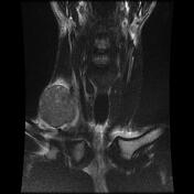
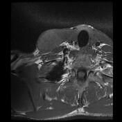
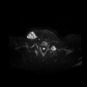
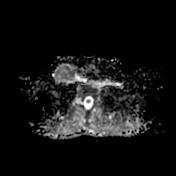
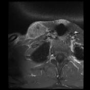
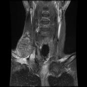
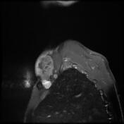

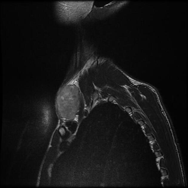
Well-circumscribed ovoid right lower cervical mass, posterolateral to the SCM muscle and anterior to the anterior scalenus muscle. It elicits an iso-to high signal to muscles on T1 and an inhomogeneous high signal on T2 with areas of restricted diffusion on DWI/ADC. The postcontrast sequences show a heterogeneous enhancement with central areas of necrosis and enhanced peripheral capsule.
No cervical lymphadenopathy was seen.
Case Discussion
The patient went on to have a complete surgical resection of the lesion with a histopathological exam that confirmed the diagnosis of clear cell sarcoma.
Clear cell sarcomas (CCS) of soft tissue are rare malignant mesenchymal tumors with melanocytic differentiation, accounting for <1% of all soft tissue neoplasms. Most frequently found in the younger adult population with women slightly more frequently affected than men.


 Unable to process the form. Check for errors and try again.