
Presentation
Abdominal pain and nausea.
Patient Data


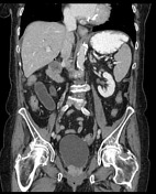
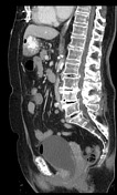
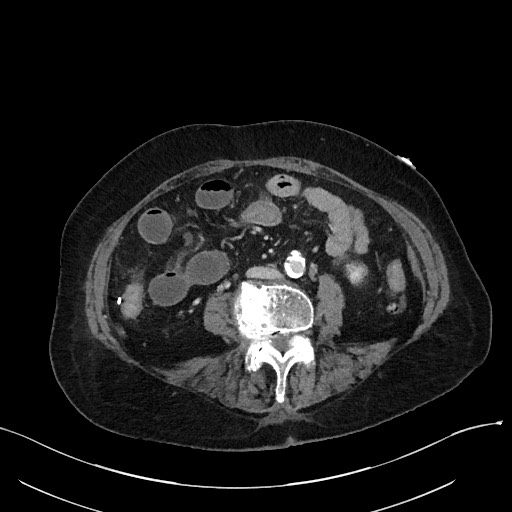
Abnormal segment of dilated, fluid-filled small bowel in the right abdomen with associated mesenteric edema/interloop fluid. Two adjacent transition points entering and exiting this segment can be seen on the same axial image, consistent with closed loop configuration. Proximal to the narrowing entering the obstruction, there is a segment of mildly dilated small bowel mid abdomen. The proximal small bowel is relatively decompressed. Abdominal aortic aneurysm. Small amount of free pelvic fluid.
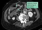
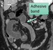
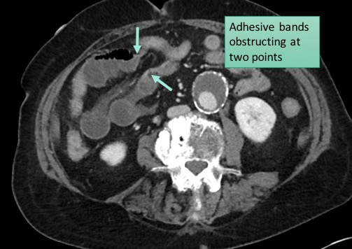
Annotated image showing buckling of the small bowel at the location of adhesive band.
Case Discussion
Adhesions are the most common cause of closed-loop obstruction. Internal hernia is the second most common cause. See further discussion on my two companion cases:


 Unable to process the form. Check for errors and try again.