
Patient Data
Note: This case has been tagged as "legacy" as it no longer meets image preparation and/or other case publication guidelines.
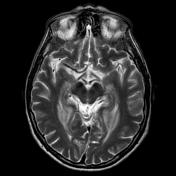

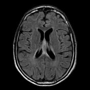
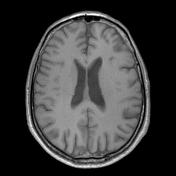
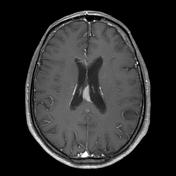
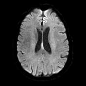
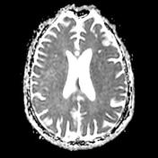
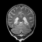

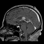
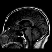
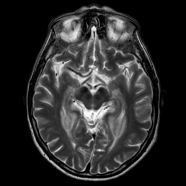
Multiple periventricular regions of vivid contrast enhancement, which are low on T2 and surrounded by relatively little edema. They also demonstrate restricted diffusion in keeping with a highly cellular tumor. Features are characteristic of CNS lymphoma.
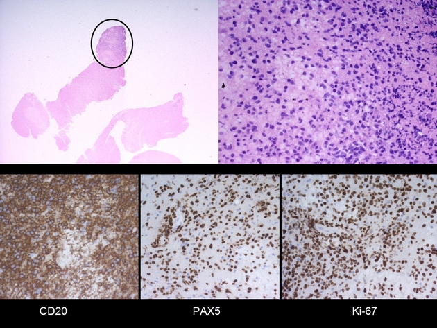
MICROSCOPIC DESCRIPTION:
The sections show a densely cellular malignant tumor in the white matter. It forms diffuse sheets and cuffs around the blood vessels. The tumor cells are intermediate in size. They have enlarged clefted and hyperchromatic nuclei, prominent nucleoli and scanty cytoplasm. The tumor cells are CD20, PAX-5, bcl-2, bcl-6 and MUM1 positive. The Ki-67 index is about 80%. They are CD3 and CD10 negative. There is insufficient tissue for EBER-CISH. The features are those of diffuse large B-cell lymphoma with activated B-cell-like phenotype.
DIAGNOSIS:
Diffuse large B-cell lymphoma.
Case Discussion
This is a typical radiological appearance of CNS lymphoma, and the diagnosis can be suggested with a high degree of certainty.


 Unable to process the form. Check for errors and try again.