
Patient Data
Age: 70 years
Gender: Male
Note: This case has been tagged as "legacy" as it no longer meets image preparation and/or other case publication guidelines.
From the case:
CNS lymphoma - steroid response

Download
Info
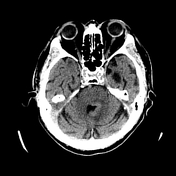
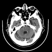
Mass lesion adjacent to the fourth ventricle, which is hyperdense on noncontrast imaging with vivid postcontrast enhancement. Adjacent hypodensity likely representing vasogenic edema.
Download
Info

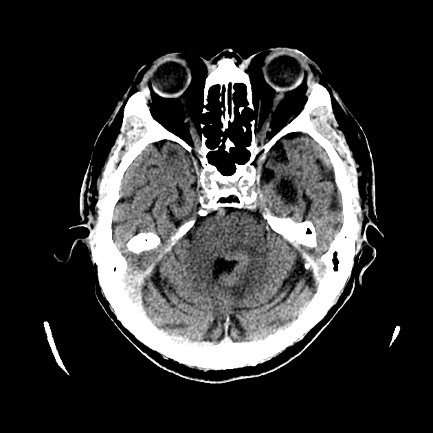
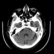
Almost complete resolution of the enhancing periventricular mass.
Case Discussion
This 70 year old man was treated with steroids on the presumptive diagnosis of CNS lymphoma. The mass is strikingly periventricular and hyperdense on precontrast CT.
On images one week later the mass has almost completely disappeared, which is characteristic of lymphoma.


 Unable to process the form. Check for errors and try again.