
Patient Data
Gender: Female
From the case:
Compartments assessed on pelvic floor MRI
Download
Info
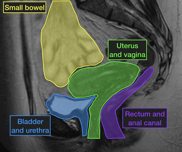
Mid-sagittal plane pelvic image showing the various compartments visible and assessed during pelvic floor MRI.
Case Discussion
Due to the soft tissue imaging capabilities of MRI, multiple compartments in the pelvis and lower abdomen can be scrutinised - analyzing the same structures using fluoroscopy requires the use of oral contrast for the small bowel, bladder contrast and contrast within the vagina, and therefore the collection of similar information on fluoroscopy is more invasive. Both MRI and fluoroscopic assessment of the posterior compartment require contrast media.


 Unable to process the form. Check for errors and try again.