Presentation
Primary infertility.
Patient Data
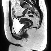

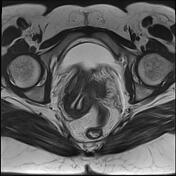

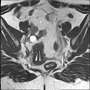

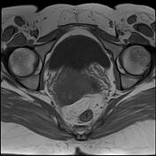


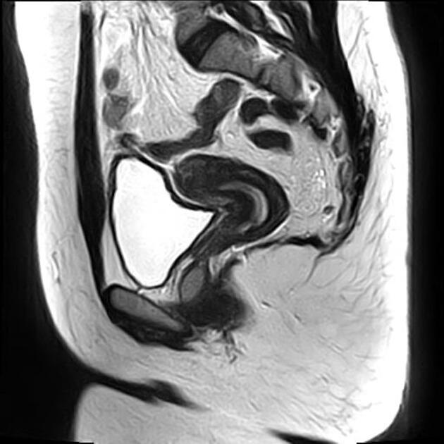
The uterus is normal in size anteverted anteflexed with two uterine cavities separated by a septum of low signal on T2, extending into endocervical canal. Mildly concave shape of the fundus surface with an acute angle between uterine cavities (35° in this case).
Bilateral ovarian follicles are the largest on the right (2 cm).
Small cyst of the right vaginal wall with a high signal on T1 (probably proteinaceous content) and an intermediate to high signal on T2 (Gartner duct cyst).
Case Discussion
MRI features of a complete septate uterus.
Septate uterus is considered the most common uterine anomaly (accounts for ~55% of such anomalies). It is classified as a class V Mullerian duct anomaly and associated with reproductive failure (67%), affecting ~15% of women with recurrent pregnancy loss.


 Unable to process the form. Check for errors and try again.