
Presentation
Delayed milestones and hypotonia
Patient Data
Age: 4 years
Gender: Male
From the case:
Congenital muscular dystrophies - cerebral manifestations


Download
Info

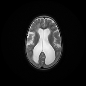
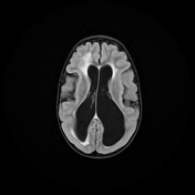
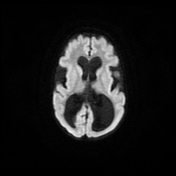
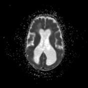
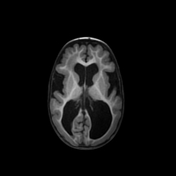
Both cerebral hemispheres show the following:
- bilateral frontal polymicrogyria
- bilateral occipital cobblestone lissencephaly
- diffuse white matter increased signal, impressive of dysmyelination.
- marked ventriculomegaly
- diffusely thinned corpus callosum
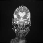
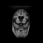
The posterior fossa shows:
- Z-shaped/kinked brainstem.
- hypoplastic pons with central ventral notching
- both cerebellar hemispheres appear small and dysplastic showing tiny cysts and abnormal cerebellar foliation
Case Discussion
The combined clinical features of hypotonia since birth, delayed motor development and the classic radiological features in this case were typical for congenital muscular dystrophy, which was confirmed on muscle biopsy.
Radiographic features
Described features in general include:
- polymicrogyria usually in the medial anterior frontal lobes
- cobblestone lissencephaly usually in the occipital lobes
- myelination defects
- ventriculomegaly
- dysgenesis of the corpus callosum
- Z-shaped/kinked, hypoplastic brainstem
- small pons
- cerebellar dysplasia, polymicrogyri with small cysts
- hypoplastic vermis


 Unable to process the form. Check for errors and try again.