
Presentation
No symptoms. Routine cardiac ultrasound screening revealed unexpected findings.
Patient Data
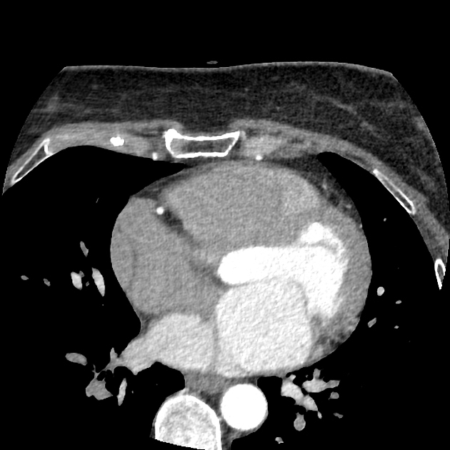
Careful examination revealed cor triatriatum. A fibrous membrane divides the left atrium into a pre-chamber receiving the pulmonary veins and a post-chamber connected to the appendage and the mitral valve. The membrane shows a large defect.
Chiari's network is in the right appendage.
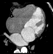
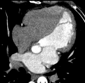

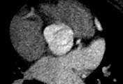

The 4-chamber view shows the fibrous membrane within the left atrium with the large communicating defect further upwards also visible in the short axis view.
The vertical long axis allows another view of the pre-chamber and post-chamber.
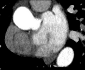
A bicuspid aortic valve, type 0 (secondary screenshot) without coarctation.
Case Discussion
Cor triatriatum is a rare congenital anomaly which may be symptomatic if the defect is narrow and obstructs blood flow through the left atrium. In this case, a large opening in the membrane allowed this middle-aged patient to remain asymptomatic.


 Unable to process the form. Check for errors and try again.