
Presentation
Seizures.
Patient Data
Note: This case has been tagged as "legacy" as it no longer meets image preparation and/or other case publication guidelines.

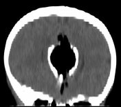
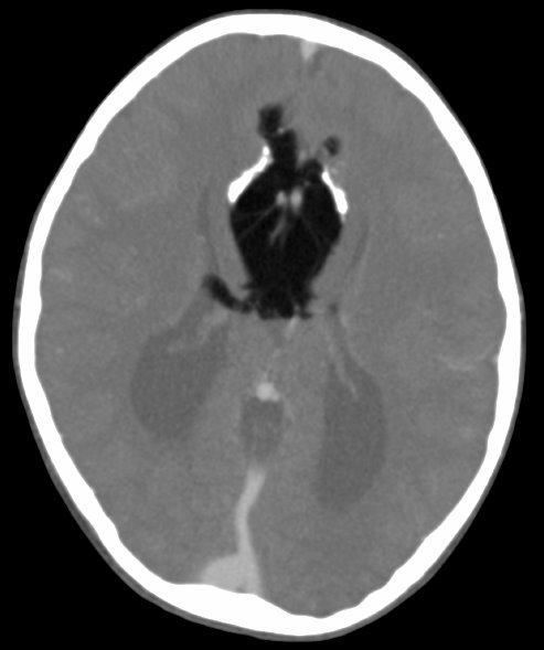
Axial post contrast CT demonstrates a fat density mass located in the interhemispheric fissure, bounded laterally by curvilinear calcifications (known as the bracket sign), best seen on the coronal reconstruction (unfortunately relatively coarse due to thick axial acquisition).
The ventricles are widely spaced with colpocephaly (racing car sign) in keeping with corpus callosal dysgenesis.


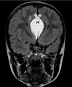
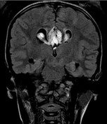
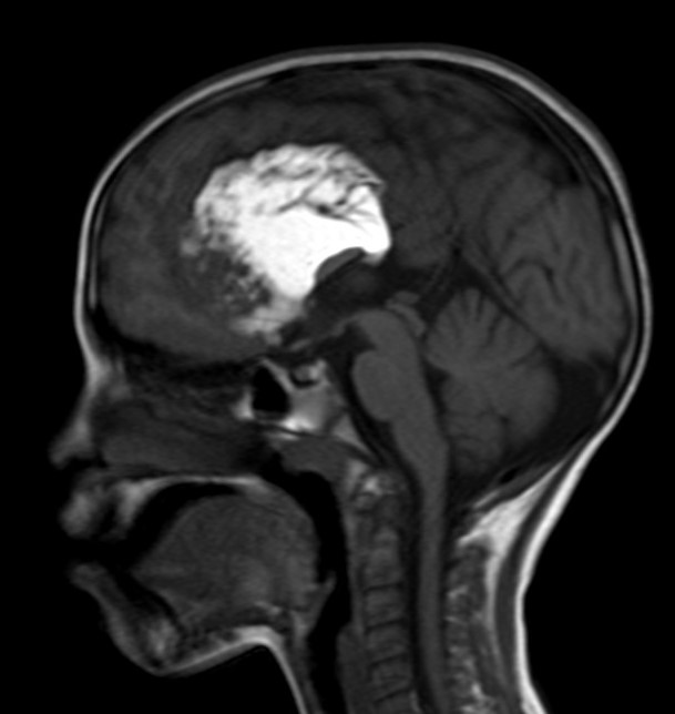
Selected images from an MRI of the brain demonstrate dysgenesis of the corpus callosum with an inter-hemispheric fatty mass entering the lateral ventricles which have a viking helmet or moose head configuration. The anterior cerebral artery and branches can be seen coursing through the superior aspect of the mass.
Case Discussion
This case demonstrates corpus callosal dysgenesis associated with a pericallosal lipoma (tubulonodular type).


 Unable to process the form. Check for errors and try again.