
Presentation
Crashed into opening car door while biking and subsequently fell on right side. No loss of conscience. Trauma protocol was initiated, consisting of conventional radiographs of chest and pelvis, followed by CT of the neck, chest and abdomen.
Patient Data
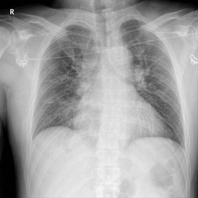
No visible trauma, in particular no fractures, lung contusions or widening of the superior mediastinum.
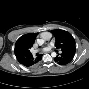

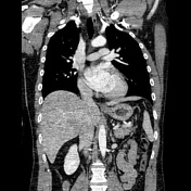
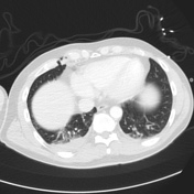
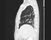


CT also shows no fractures and no lung contusions. However, there is a distinct fracture of the costosternal cartilage at the level of the 4th through 7th rib on the right with herniation of lung tissue through the defect. Interestingly, no pneumothorax is demonstrated, suggesting the pleura is still intact and herniates together with the lung.
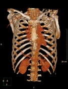
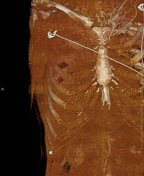
The cartilage fracture can be easily overlooked when focusing only on bony fractures or lung tissue and doesn't show up in the commonly used bone window 3D volume rendering.
No other injuries were noted.
Case Discussion
Primary complaint was pain, no dyspnea. Patient went home with painkillers. Although dislocation was noticeable during physical examination on subsequent outpatient clinic visits, pain decreased and patient was discharged.
Costal cartilage fractures are uncommon and undetectable on plain radiographs, unless the cartilage is calcified. CT, ultrasound and MRI can all demonstrate fractures of the costal cartilage. In a pediatric population and in follow-up examination ultrasound may be preferable.
These fractures are commonly left untouched, although it remains unknown whether the cartilage fully heals.


 Unable to process the form. Check for errors and try again.