Presentation
Fever, associated cough, and shortness of breath (dyspnea ++) for a week. He denies a sore throat.
Patient Data


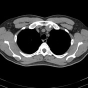
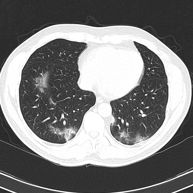
CT demonstrates multilobar and bilateral ground-glass opacities with rounded morphology, mostly in the periphery of both lungs.
There is no mediastinal hilar or axillary lymphadenopathy, nor pleural/pericardial effusion.
Findings are typical of covid-19 pneumonia.
Case Discussion
This patient tested positive for coronavirus disease-19.
Coronavirus disease-19 or COVID-19 is a viral infectious disease caused by SARS-CoV-2 1. A combination of chest imaging elements and repeat laboratory RT-PCR may help to increase the COVID-19 diagnosis 1,2,3. This patient had laboratory-confirmed SARS-CoV-2, and he had the typical CT features of COVID-19 pneumonia. The patient recovered well.


 Unable to process the form. Check for errors and try again.