
Presentation
Admitted to ED with shortness of breath
Patient Data
Age: 70 years
Gender: Female
From the case:
COVID-19 pneumonia
Download
Info

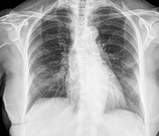
There is a coarsening of lung markings more evident at the lower fields (R>L) but no clear consolidation seen.
Surgical clips overlie the right breast shadow.
From the case:
COVID-19 pneumonia


Download
Info
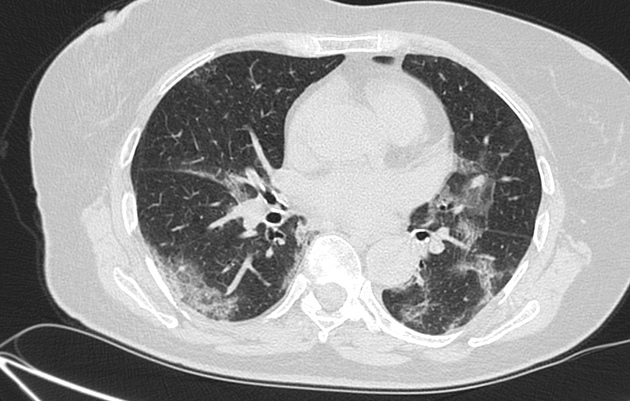
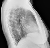
Bilateral ground-glass opacities are seen in both lungs, mostly mid to lower zones.
Non-specific mediastinal lymph nodes.
Surgical clips at the right breast.
Case Discussion
This patient tested positive for COVID-19, without a travel history to high-risk areas (including Northern Italy).
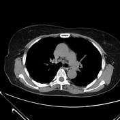
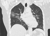
Chest CT appearance is almost typical, including ground-glass opacities, air-space opacification and crazy paving with no significant lymphadenopathy.
Further reading (external links)
- latest articles on COVID-19 - European Radiology
- special focus: COVID-19 - RSNA journals


 Unable to process the form. Check for errors and try again.