
Presentation
Recent travel from endemic COVID-19 region. 24 hours confusion with new temperature and desaturation on assessment.
Patient Data
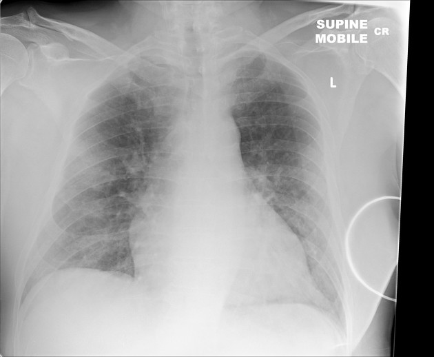
Supine study. Patchy consolidation in peripheral right midzone.
No pleural abnormality.
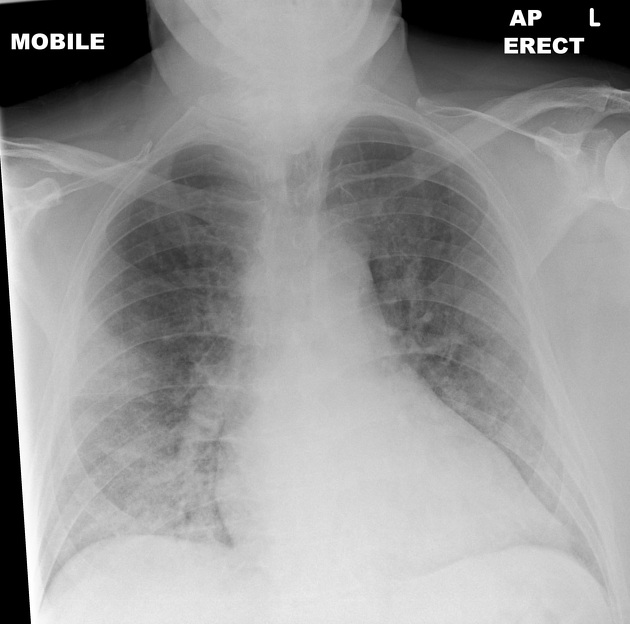
Progressive right mid and lower zone consolidation. Left lung and pleural spaces remains clear.
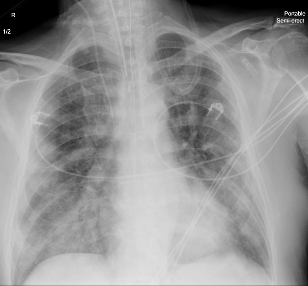
Diffuse patchy airspace consolidation in the mid/lower zones bilaterally.
ET tube, right jugular central line and NG tube in situ.
No pleural abnormality.


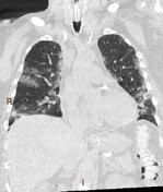
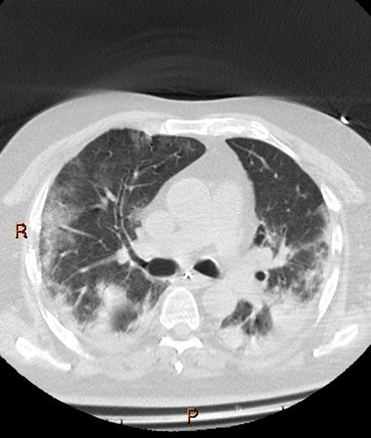
Day 13 performed to assess degree of lung injury
Multifocal regions of consolidation and ground-glass opacifications.
These have a peripheral and basal predominance.
No pleural or pericardial effusion.
Case Discussion
Serial imaging demonstrating progressive changes in a patient with PCR confirmed COVID-19 infection.
There can be a rapid deterioration in imaging findings.
Please read the COVID-19 article linked below for further information and examples.


 Unable to process the form. Check for errors and try again.