
Presentation
Fever and cough for a few days.
Patient Data
Age: 80
Gender: Male
From the case:
COVID-19 pneumonia


Download
Info
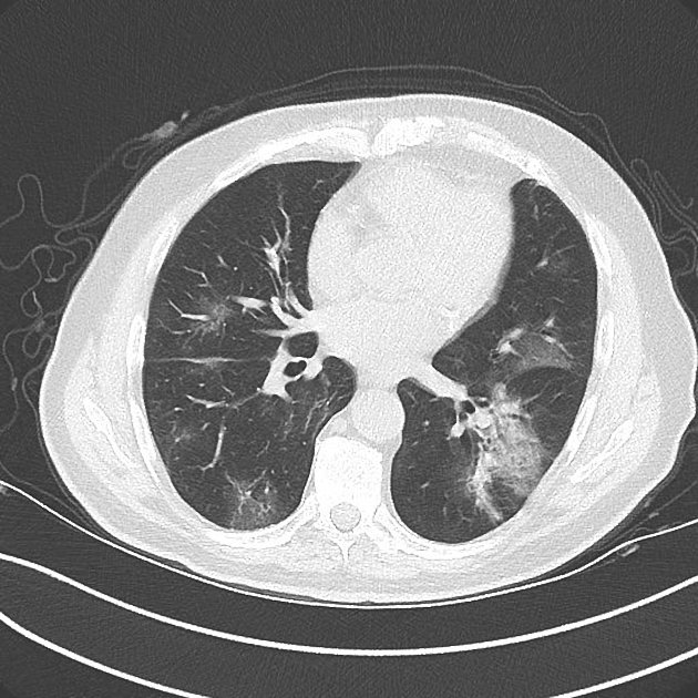
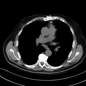
Axial CT shows multilobar ground-glass opacities with peripheral and mid to basal lobe predominance. Airspace consolidation in the left lower lobe.
No significant mediastinal lymphadenopathy.
Incidental pleural nodular calcific plaques.
Previous coronary bypass surgery.
Case Discussion
The RT-PCR COVID-19 test was positive. This patient was considered to have COVID-19 pneumonia.


 Unable to process the form. Check for errors and try again.