
Presentation
Fever, cough and chest pain from 5 days ago.
Patient Data


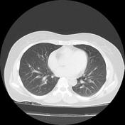
Ground glass nodule is present at the left lower lobe laterobasal segment.
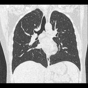

Ground glass nodule is present at the left lower lobe laterobasal segment.
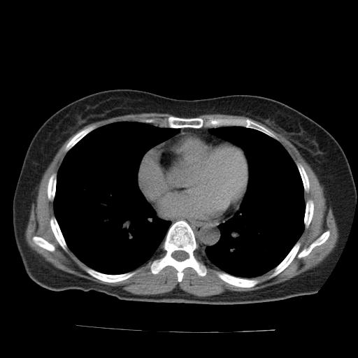
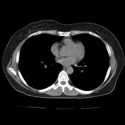
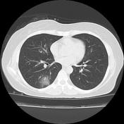
Peripheral ground-glass opacities are seen at the right lower lobe superior and basal segments as new findings.
Case Discussion
The interesting point of the recent case is in the delay imagining presentations of COVID-19 pneumonia.
When the patient referred to our imaging center for chest CT, we saw only a small ground-glass nodule at the left lower lobe. The patient didn't have a previous CT for comparison, because according to our experiences almost COVID-19 pneumonia will be detected at least on the fifth day after initial presentations.
So regarding the clinical presentations and high prevalence of COVID-19 pneumonia in Iran, we repeat the chest CT, 48 hours later and finally, we found the new ground-glass opacities in the right lower lobe.
Subsequently, the patient underwent RT-PCR COVID-19 assay and the result was positive.


 Unable to process the form. Check for errors and try again.