
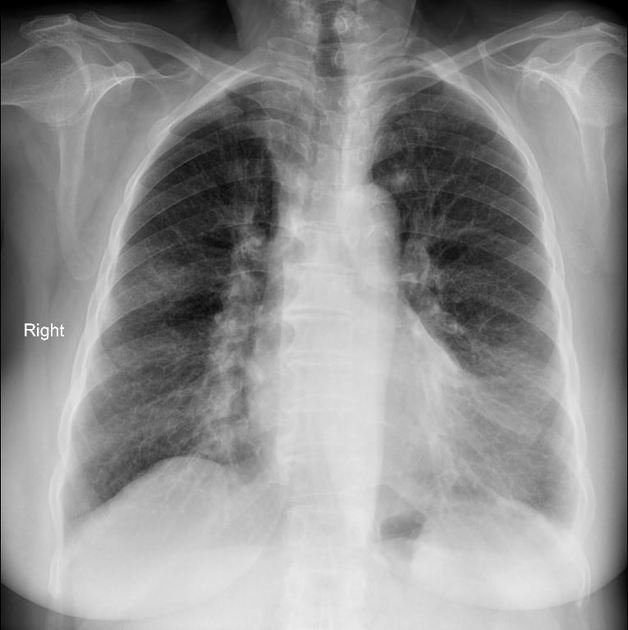
Presentation
Fever and cough.
Patient Data
Age: 60 years
Gender: Female
From the case:
COVID-19 pneumonia
Download
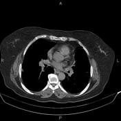
Info
Patchy ill defined subpleural opacities are seen particularly at mid zone of right lung.
From the case:
COVID-19 pneumonia


Download
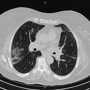
Info
Bilateral subpleural illdefined ground glass opacities are seen more prominent at upper lobes. Subpleural ill-defined consolidations are noted at posterior segment of right upper lobe. In addition, partial collapse is noted at left lingular segment. There are also a few atelectatic bands at lung bases.
Case Discussion
This patient had positive RT-PCR testing for COVID-19.


 Unable to process the form. Check for errors and try again.