
Presentation
Fever, cough, breathing difficulties for about ten days.
Patient Data
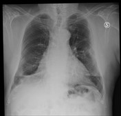
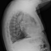

Vertical air space consolidation along the left costal margin.
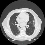

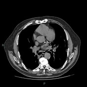
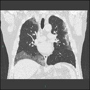


There are large areas of ground glass opacities in the lower right lobe, in the upper lobes, with interlobular septal thickening in the subpleural area. Paraseptal emphysema is present in the upper lobes. No evidence of mediastinal adenopathy.
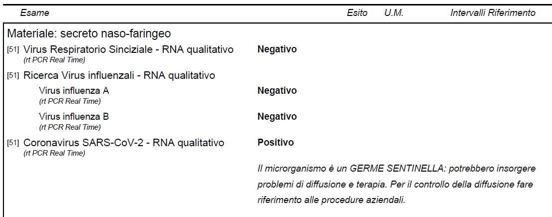
Cytological report showing positivity for SARS-CoV-2 in a nasopharyngeal aspirate using the confirmatory reverse transcriptase-polymerase chain reaction (RT-PCR) test.
Case Discussion
In the right clinical context large ground-grass opacity lesions, predominantly in the peripheral and posterior lungs on CT, are diagnostic of COVID-19 pneumonia. Note the absence of mediastinal adenopathy which is not usually seen in COVID-19 and should suggest the presence of superadded infection, e.g. bacterial pneumonia.
Case courtesy: Dr ssa Sandra Pennacchini


 Unable to process the form. Check for errors and try again.