Q: May chest CT be diagnostic in some cases with an initial negative RT-PCR?
show answer
A: CT findings may be diagnostic in many cases with an initial false-negative RT-PCR screening test. A combination of chest imaging elements and repeat laboratory RT-PCR may help to increase the COVID-19 diagnosis.
Q: Why is chest CT useful in patients with suspected COVID-19?
show answer
A: CT is recommended in suspected COVID-19 cases because of the primary involvement of the respiratory system. CT findings may occur even before symptom onset.
Q: What are the primary findings of COVID-19 on chest computed tomography?
show answer
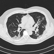
A: Many patients have unremarkable CT scans within two days of symptom onset. So, a normal chest CT examination does not exclude the infection. The typical CT features of COVID-19 on initial CT are bilateral, multilobar ground-glass opacities (GGO), sometimes with a rounded morphology and a peripheral and basal lung predominant distribution. Air space consolidations superimposed on GGO may occur in this stage, mainly in the elderly population. In the intermediate stage of the disease, usually, there is an increase in the size and number of GGOs and the transformation of GGO into multifocal consolidative opacities, septal thickening, and development of a crazy-paving pattern, which consists of GGO and inter/intralobular septal thickening. The highest severity of CT findings occurs around day ten after the symptom onset. CT signs of clinical improvement usually occur after two weeks of the disease, which are the gradual resolution of consolidative opacities and a decrease in the number of lesions and involved lobes.
Q: Which are some uncommon CT findings of COVID-19?
show answer
A: Some uncommon CT findings of COVID-19 are air bronchogram, bronchiectasis, pleural thickening, and subpleural involvement thickening mainly in the later stage of the infection. Other uncommon findings include Pleural effusion, pneumothorax, CT reverse sign, halo sign, cavitation, lymphadenopathy, and pericardial effusion, which may occur with disease progression.
Q: What are the chest CT findings that have the highest discriminatory value?
show answer
A: The following CT findings have the highest discriminatory value: ground-glass opacities (GGO) with or without consolidations and crazy-paving appearances composed by GGO with reticular and/or interlobular septal thickening with predominant bilateral, multifocal and peripheral distribution. In some cases, there is a simultaneous presence of these patterns in the same patient, suggesting a progression of pulmonary involvement.


 Unable to process the form. Check for errors and try again.