
Presentation
Shortness of breath, fever, cough.
Patient Data
Age: 70 years
Gender: Female

Download
Info

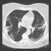
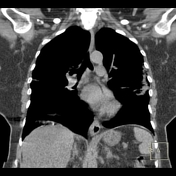
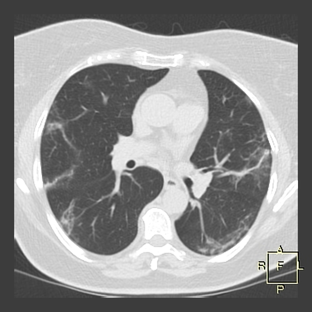
Bilateral subpleural ground-glass opacity, linear subpleural consolidation in left upper lobe (LUL).
No pleural effusion.
No mediastinal lymphadenopathy.
This patient has had positive RT-PCR testing for 2019-nCoV before CT, so CT findings are consistent with COVID-19 pneumonia CO-RADS 6.

Download
Info
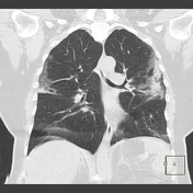
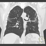
Transforming ground-glass opacity to subpleural fibrous stripes.
Case Discussion
Initial chest CT shows typical radiological signs of COVID-19 pneumonia. Next chest CT shows an absorption stage of COVID-19 pneumonia.


 Unable to process the form. Check for errors and try again.