
Presentation
One week history of fever, dysuria. Presents to the ER with abdominal pain and nausea. No respiratory symptoms. Fever and elevated CRP. Hyperlipidemia and hypertension.
Patient Data


Findings in the abdomen consist of gallstones without evidence of cholecystitis, a liver cyst in segment 7 and an unclear focal liver lesion in segment 4A and a lesion in the right adrenal gland. All findings were present at a CT-colongraphy done several month earlier. In that study (with and without i.v. contrast) the adrenal lesion was consistent with a lipid-rich adenoma.
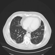
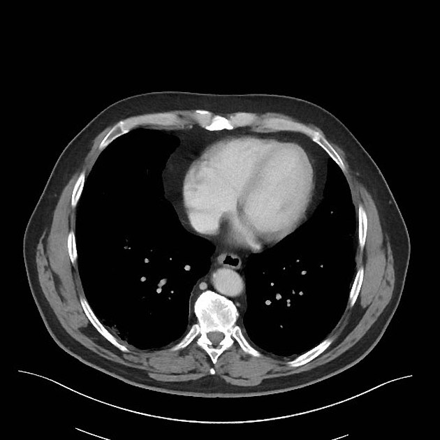
Additional findings: Patchy ground-glass opacities in basal lungs bilaterally.
Case Discussion
The radiologist suggested COVID-19 and subsequent testing was positive for SARS-CoV-2 virus.
There are many case reports of SARS-CoV-2-infected patients presenting with only abdominal symptoms and fever, without symptoms from the respiratory tract.


 Unable to process the form. Check for errors and try again.