
Presentation
Fever, cough and breath shortness in a known patient with old tuberculosis.
Patient Data
Age: 65 years
Gender: Male
From the case:
COVID-19 pneumonia in a patient with old tuberculosis


Download
Info

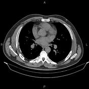
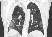
Patchy ill-defined opacities are seen at both lungs particularly at subpleural and basal regions.
Focal subpleural fibrotic opacity is also noted at right lung apex. In addition, multiple tiny calcified nodules, less than 4 mm are evident bilaterally. Pleural thickening and calcification are seen at right posterolateral aspect of right hemithorax.
Mild degenerative changes as osteophytes are seen at the thoracic spine.
In imaged portions of upper abdomen, two stones 12 mm and 10 mm are present in gallbladder.
Case Discussion
This patient had positive RT-PCR testing for COVID-19.


 Unable to process the form. Check for errors and try again.