
Presentation
Routine CT thorax/abdomen follow up for oncologic treatment of a GI tract cancer. No known metastases. The patient was clinically well.
Patient Data


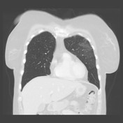

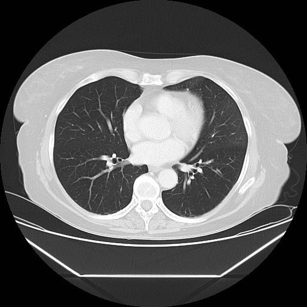
Peripheral ground glass opacifications with diffuse delineation in the left upper lobe, not present on previous exams. No other parenchymal changes. No lymphadenopathy, no pleural effusions.
Case Discussion
This incidental finding was reported to the clinician as suspicious for COVID-19, and subsequent PCR testing confirmed the diagnosis.
The case serves to illustrate that even patients presenting with no symptoms may have changes on chest CT. This is in keeping with previous reports, e.g. from the Diamond Princess, where 54% of asymptomatic but PCR confirmed cases proved to have CT findings 1.


 Unable to process the form. Check for errors and try again.