
Presentation
Short of breath. No history of respiratory disease.
Patient Data
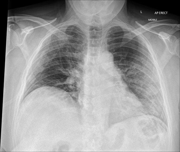
Minor bilateral mid zone airspace opacification in a peripheral distribution.
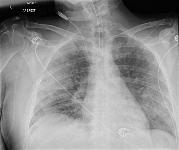
Endotracheal tube and right internal jugular lines.
Unchanged airspace opacification in the lungs.

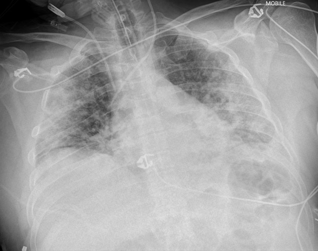
Endotracheal tube, right internal jugular line and nasogastric tubes suitably sited.
Bilateral peripheral airspace opacification which has progressed since the prior radiograph.
No pleural effusions.
ET tube and bilateral internal jugular lines.
Further progression in the bilateral airspace opacification with a more peripheral distribution than on the prior radiograph.
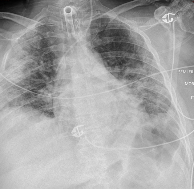
Tracheostomy and left internal jugular line.
Minor regression of the left sided airspace opacification.
Case Discussion
This is a series of chest radiographs to demonstrate the longitudinal imaging appearances of COVID-19.
As in this case, a significant number of long stay ICU patients end up with tracheostomy insertion.


 Unable to process the form. Check for errors and try again.