
From the case:
Coxa valga
Download
Info
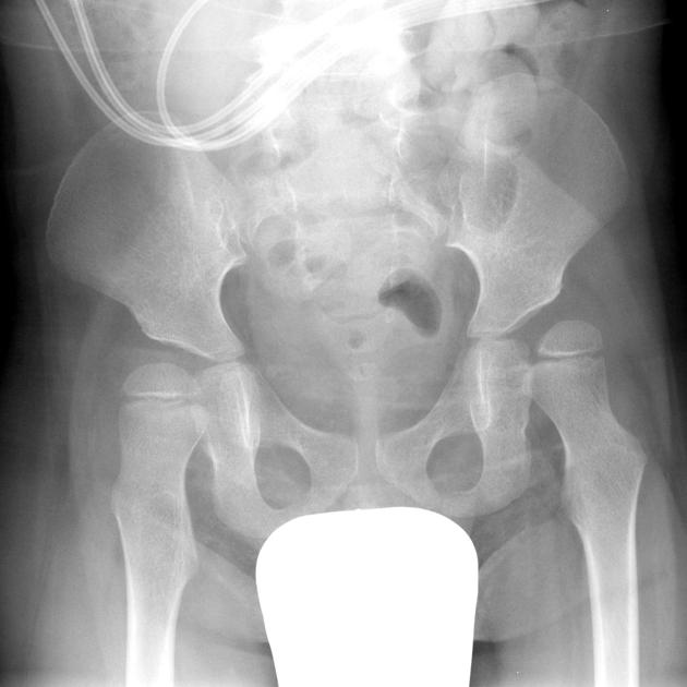
Single view of the pelvis and both hips demonstrates bilateral reduced femoral neck angle resulting in lateral subluxation of the femoral head out of the acetabulum.
Case Discussion
This case illustrates bilateral coxa valga with resultant subluxation. It should not be mistaken for DDH.


 Unable to process the form. Check for errors and try again.